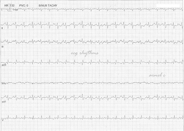
What
is your ECG interpretation?
Figure
1 - ECG case in full disclosure
Figure
2 - P waves marked
This
is a regular wide QRS complex tachycardia at a rate of about 130's bpm with a
right bundle branch block morphology.
There
are 2 sets of P waves that can be appreciated. The first p wave morphology (red
arrows) has a cycle of about of 440 ms or at 136 bpm. The second P wave
morphology (blue arrows) has a cycle of about 560 ms or at 107 bpm. The second
P wave morphology is sometimes hidden from view or are buried in the QRS (black
arrows). Occasionally, fusion of the P waves can be seen (green arrows and F).
Both P waves are inverted in aVR. The
two P waves are dissociated. So, we have atrial dissociation with the other P
wave conducted with 1:1 ratio with a RBBB morphology.
This
ECG is a from a post-cardiac transplant patient. Two sets of P waves frequently
results from a procedure that sutures the donor heart to the corresponding
structures of the recipient residual atria. The 2 sets of P waves are usually
dissociated from each other but in some cases becomes synchronized. Unusual
atrial rhythms also develop particularly when one of the sets of the 2 atrial
components develops ectopic activity, atrial fibrillation or atrial flutter. A
complete or incomplete RBB is present in more than 80% of ECGs after
transplantation.
Reference:
Surawicz
B and Knilans TK. 2008. Chou’s Electrocardiography in Clinical Practice. 6th
ed. PA. Saunders-Elseiver
#657


No comments:
Post a Comment
Note: Only a member of this blog may post a comment.