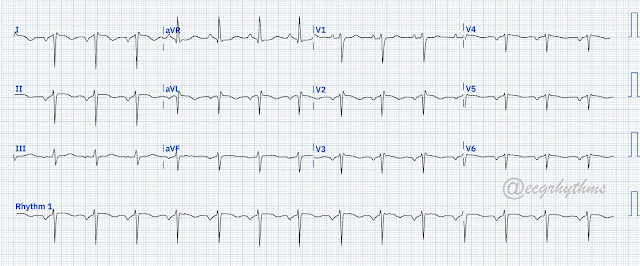
No clinical hx. What is your interpretation?
Image Case (Digitized using PMCardio App)
This is a regular narrow QRS complex rhythm with inverted P waves in I, inferior leads, V2-V6 and upright in aVR with PRI of around 0.16 seconds. There is low QRS voltage in V4-V6. The leads are in the correct location. This is dextrocardia (Image 2) with ectopic atrial rhythm. P waves that are in the superior direction (ectopic atrial pacemaker) can be seen in patients with polysplenia syndrome (noted on this patient).
Image 2 - Chest x-ray
Reference:
Park M. 2016. Park's Pediatric Cardiology Handbook. 5th ed. PA Elsevier
Surawicz B and Knilans TK. 2008. Chou’s Electrocardiography in Clinical Practice. 6th ed. PA. Saunders-Elseiver
#683


No comments:
Post a Comment
Note: Only a member of this blog may post a comment.